
File:Fundus photograph of normal right eye.jpg

預覽大小:600 × 600 像素。 其他解析度:240 × 240 像素 | 480 × 480 像素 | 768 × 768 像素 | 1,024 × 1,024 像素 | 1,411 × 1,411 像素。
原始檔案 (1,411 × 1,411 像素,檔案大小:248 KB,MIME 類型:image/jpeg)
摘要
| 描述 |
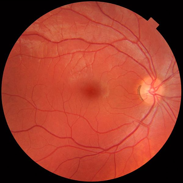
English: Fundus photograph of the right eye, showing a fundus with no sign of disease or pathology. It is seen from front so that left in each image is to the person's right. The gaze is into the camera, so the macula is in the center of the image, and the optic disk is located towards the nose (right in image). The optic disk has some pigmentation at the perimeter of the lateral side, which is considered non-pathological.
Veins are darker and slightly wider than corresponding arteries. Major nerve pathways are seen as white striped patterns radiating from the optic disk. In addition, there are also lighter areas close to larger vessels seen mainly at upper left in the image (person's upper right), which is regarded as a normal finding in younger people. Photo is taken at Gävle Hospital in Sweden in 2012 on a healthy 25-year old male volunteer. |
| 日期 | |
| 來源 | 自己的作品 |
| 作者 |
When using this image in external works, it may be cited as:
or
|
| 其他版本 |
 |
授權條款
我,本作品的著作權持有者,決定用以下授權條款發佈本作品:
| 此檔案在創用CC CC0 1.0 通用公有領域貢獻宣告之下分發。 | |
| 在此宣告之下分發本作品者,已依據各國著作權法,在全世界放棄其對本作品所擁有的著作權及所有相關相似的法律權利,從而將本作品貢獻至公有領域。您可以複製、修改、分發和演示該作品,用於任何商業用途,所有這些都不需要請求授權。
|
檔案歷史
點選日期/時間以檢視該時間的檔案版本。
| 日期/時間 | 縮圖 | 尺寸 | 用戶 | 備註 | |
|---|---|---|---|---|---|
| 目前 | 2012年3月21日 (三) 10:49 | | 1,411 × 1,411(248 KB) | Mikael Häggström |
檔案用途
下列頁面有用到此檔案:
全域檔案使用狀況
以下其他 wiki 使用了這個檔案:
- ar.wikipedia.org 的使用狀況
- bs.wikipedia.org 的使用狀況
- ca.wikipedia.org 的使用狀況
- de.wikipedia.org 的使用狀況
- en.wikipedia.org 的使用狀況
- en.wikiversity.org 的使用狀況
- et.wikipedia.org 的使用狀況
- fr.wikipedia.org 的使用狀況
- fr.wikiversity.org 的使用狀況
- gl.wikipedia.org 的使用狀況
- he.wikipedia.org 的使用狀況
- hu.wikipedia.org 的使用狀況
- it.wikipedia.org 的使用狀況
- ml.wikipedia.org 的使用狀況
- pl.wikipedia.org 的使用狀況
- simple.wikipedia.org 的使用狀況
- th.wikipedia.org 的使用狀況
- tr.wikipedia.org 的使用狀況
- uk.wikipedia.org 的使用狀況
- zh-yue.wikipedia.org 的使用狀況